If it has been a while since your last real eye exam, it is fair to wonder what you are actually signing up for. Is it the letter chart and a new pair of glasses? Is it the puff of air? Do they have to dilate you? And how much of your afternoon does it take?
A comprehensive eye exam typically includes your health history, visual acuity, refraction, eye-movement and pupil testing, eye-pressure measurement, a slit-lamp exam of the front of the eye, and an evaluation of the retina and optic nerve — usually with dilation — plus visual-field and color testing as indicated. That is the American Optometric Association’s adult component list. A visit commonly runs 45 to 90 minutes. Jackson Davenport Vision Center performs comprehensive exams in Summerville.
Here is what a full visit covers, component by component, and what each part is looking for. Some of it happens at every exam; some of it is added when your history, symptoms, or risk call for it.
The Standard Behind the Exam
The exam is not improvised. The American Optometric Association (AOA) publishes an evidence-based clinical practice guideline for adults, listed on its clinical practice guidelines portal as Comprehensive Adult Eye and Vision Examination, Second Edition. Its current version reflects a 2023 update — the revision that moved the AOA to recommend annual exams for adults 18 to 39. That guideline is the optometry profession’s own definition of a comprehensive adult exam: a defined set of core components, plus additional testing chosen according to your history, symptoms, age, and risk.
Two things follow from that. First, a comprehensive exam is a medical evaluation of your eyes, not just a prescription check. Second, because the doctor can see your blood vessels and optic nerve directly, the exam can turn up early signs of conditions elsewhere in the body — including diabetes and high blood pressure — that a patient has no way to feel yet.
The Sequence: History, Acuity, and the Refraction That Sets Your Prescription
Your history
Every exam starts with questions, and they are not small talk. Your personal, family, and medication history tells the doctor where to look hardest — a parent or sibling with glaucoma, a diabetes diagnosis, a history of eye injury, or a medication that requires retinal monitoring all change what the rest of the visit needs to cover.
Visual acuity
Next comes the familiar letter chart, measuring how sharply you see at distance and up close. “20/20” comes from this test. Acuity is a useful measure of the eye’s resolving power, and a drop in it can be an early sign of a refractive change or of an underlying problem such as cataract or macular disease — which is exactly why the number gets recorded and compared year over year.
Refraction: objective, then subjective
Setting your prescription usually takes two passes.
- Objective refraction uses an instrument — a retinoscope or an autorefractor — to measure how light bends as it enters your eye. It needs no answers from you at all, and it gives the doctor a measured starting point to refine.
- Subjective refraction is the part you know: “Which is clearer, one or two?” When it is appropriate, your responses refine that starting point into what actually looks and feels best to you.
Together they land the correction for nearsightedness (myopia), farsightedness (hyperopia), and astigmatism. If you are past about age 40 and near work has gotten harder, this is also where presbyopia — the normal, age-related stiffening of the eye’s natural lens — shows up.
Motility, pupils, side vision, and color
Several quick tests round out the functional half of the exam, and each one has a specific job:
- Ocular motility evaluates how your eyes move and whether they stay aligned, which reflects how well the eye muscles are working.
- Pupil testing checks how your pupils respond to a bright light. Pupils normally get smaller; a response that is sluggish or unequal is something the doctor follows up on.
- Side vision is checked by confrontation or with automated visual-field testing, as indicated. Loss of peripheral vision can be a symptom of glaucoma, and it is easy to miss on your own — you can lose side vision without noticing.
- Color vision testing, when indicated, looks at how you distinguish colors. A deficiency can be something you were born with, or something acquired from an optic-nerve or macular condition.
Eye pressure
Tonometry measures the fluid pressure inside the eye, called intraocular pressure. Elevated pressure is an important, measurable risk factor for glaucoma — a disease that damages the optic nerve, usually causes no symptoms until peripheral vision is already gone, and is not curable, though it is manageable when it is found early. Pressure alone does not settle the question: glaucoma can occur without elevated pressure, so the doctor weighs the pressure reading together with the appearance of your optic nerve and your visual field. Regular exams are the only way to catch it in time. You can read more about how we handle glaucoma testing here in Summerville.
The slit lamp
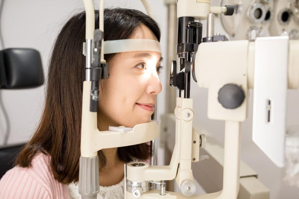
The slit-lamp biomicroscope gives a magnified, three-dimensional view of the front of your eye: eyelids, conjunctiva, cornea, iris, and the natural lens. It is how a doctor sees corneal changes, evaluates the tear film and ocular-surface problems such as dry eye disease, and looks directly at the lens for cataract — the clouding that most often comes with age and shows up as blur, glare, halos around lights, and colors that look faded.
The Part Screenings Skip: Binocular Vision, Focusing, and Eye Teaming
A quick screening asks whether you can read the chart. A comprehensive exam asks whether your two eyes work together comfortably, and whether they can hold focus at near for as long as your job demands. The AOA guideline calls for assessing ocular motility, binocular vision, and accommodation in adults. These tests are less familiar than the letter chart, and a screening generally does not provide the assessment of binocular vision and focusing that a comprehensive exam does.
The tests are simple and quick:
- Cover test and phorias — a phoria is a latent tendency for one eye to drift in or out, revealed only when the doctor briefly interrupts the two eyes working as a pair. You would not see the deviation itself, though it can show up indirectly as eyestrain, headaches, or intermittent double vision.
- Near point of convergence — the closest point at which you can hold a single image before one eye drifts outward.
- Stereopsis — depth perception, which confirms your brain is fusing both images into one three-dimensional picture.
- Accommodative amplitude and facility — the focusing power your eyes can produce, and how quickly they can shift focus between near and far. (Focusing power declines naturally with age; the facility test uses a small handheld tool with alternating lenses.)
Why bother, if you see 20/20? Because an adult can have perfect acuity and still have a convergence or focusing problem. Those problems tend to show up as eye strain, headaches across the front of the head, intermittent double vision, fatigue during computer work, or losing your place while reading. None of that is a diagnosis you should make about yourself from a blog post — it is a reason to have the testing done, so an eye doctor can tell you whether the eyes are the cause.
Related, and worth knowing: a lot of screen-related discomfort is plain digital eye strain from blinking less and holding focus at one distance for hours. The accepted guidance is the 20-20-20 rule — every 20 minutes, look at something at least 20 feet away for at least 20 seconds.
The Health Half: Dilation vs. Digital Retinal Imaging, and What Each Can Catch
The back of the eye — the retina, macula, optic nerve, and retinal blood vessels — is where a comprehensive exam earns its name. This is where a doctor can find age-related macular degeneration (by deposits called drusen, or by leakage in the macula), diabetic retinopathy (by tiny vessel bulges, small hemorrhages, or abnormal new vessel growth), hypertensive changes in the retinal vessels, and glaucoma damage at the optic nerve head.
There are two ways to look, and they are not interchangeable.
What dilation gives you
A dilated exam uses drops to widen the pupil so the doctor can get a wide, dynamic, three-dimensional view of the retina — out toward the far edges. That stereo view is what allows an assessment of depth and elevation at the optic nerve and macula, and it is the standard approach for finding peripheral tears, holes, and detachments.
The AOA’s adult guideline states it plainly: “pharmacologic dilation of the pupil is generally required for thorough stereoscopic evaluation of the ocular media, retinal vasculature, macula, optic nerve, and the peripheral retina.” Dilation is specifically indicated for patients with diabetes, strong nearsightedness, or a history of flashes and floaters.
One important distinction there: a long-standing, stable floater is a routine finding your doctor tracks, but a sudden shower of new floaters or flashes of light — or a curtain or shadow across part of your vision — can signal a retinal tear or detachment. Those are reasons to contact an eye doctor right away or seek emergency care, not reasons to wait for your next annual visit.
What imaging adds
Widefield digital retinal imaging captures a high-resolution photograph of a broad area of the retina in a fraction of a second, often without drops. It adds real value: a permanent record to compare against next year’s image, filters that make subtle findings easier to see, and a picture your doctor can turn around and show you.
Optical coherence tomography (OCT) is a different kind of look — a cross-sectional scan through the retinal layers that can reveal structural change in glaucoma and macular disease. The AAO lists OCT, fundus photos, and automated visual fields among the additional tests a doctor may add to a comprehensive exam.
The honest answer on “do photos replace dilation?”
No. Imaging is a companion to a dilated exam, not a substitute for one — photographs can miss the extreme edges of the retina, and a photograph is not the same as the dynamic, three-dimensional look a doctor gets through a dilated pupil. Whether you need dilation at a given visit is a clinical decision your eye doctor makes from your risk factors, symptoms, and history.
What dilation feels like, and the driving question
The drops need time to take effect before the doctor can look, which is part of why the visit runs as long as it does. Afterward, expect light sensitivity and blurry near vision — the AAO tells patients their eyes “might be sensitive to light for a few hours after dilation.” How long it lasts depends on the drops used and on you, so ask what to expect at your visit.
The simplest plan is to bring a driver or arrange a ride, and to ask us in advance if you are not sure whether you will be dilated. Dark wraparound sunglasses help with the glare on the way out, and night driving is worth avoiding entirely until the drops have worn off. If your vision is not clear enough to drive safely, do not drive.
How Often Adults Need One: The AOA and AAO Schedules, Kept Separate
This is where online answers most often go wrong. The optometry body and the ophthalmology body publish different adult schedules, and they should never be blended into a single “every one to three years” rule. Here is each, as its own position.
The AOA, in the 2023 update to its adult guideline, recommends annual comprehensive eye exams for asymptomatic, low-risk adults in every adult age band:
- 18–39 — Annually
- 40–64 — Annually
- 65+ — Annually
For patients considered at-risk, the AOA recommends exams at least annually, or more often as the doctor advises.
The AAO (American Academy of Ophthalmology), in its Comprehensive Adult Medical Eye Evaluation Preferred Practice Pattern, recommends the following for asymptomatic patients without risk factors for eye disease:
- Under 40 — Every 5 to 10 years
- 40 to 54 — Every 2 to 4 years
- 55 to 64 — Every 1 to 3 years
- 65 or older — Every 1 to 2 years
The AAO also tells adults with healthy eyes to get a baseline eye-disease screening by age 40, when early signs of disease often first appear.
Both schedules are real, and each belongs to the organization that published it. Where they agree completely is that risk changes the answer:
- Diabetes. The AAO’s practice pattern sets the timing by diabetes type: a first examination within five years of onset for type 1 diabetes and at the time of diagnosis for type 2, then at least yearly. Pregnancy changes the schedule again. Diabetic eye disease is silent early, which is the entire reason for the schedule — and your own doctor sets the interval that fits your case.
- Glaucoma risk factors. The AAO recommends more frequent evaluations for adults with risk factors for primary open-angle glaucoma, including diabetes, older age, and family history.
- Strong nearsightedness. High myopia is often associated with a longer eye, which stretches and thins the peripheral retina and raises retinal risk — a reason your doctor may want to look at the retina more often, and with dilation.
- Prior eye surgery or injury. A previous procedure or injury is another reason the interval gets set to your specific history and findings rather than to the standard schedule.
- Certain long-term medications call for their own retinal monitoring, on a schedule set by your prescriber and your eye doctor together. Bring a complete medication list to every exam and ask whether any of yours falls into that group.
The safe, true version for any individual: how often is right for you depends on your age, your health, and your risk, and your eye doctor sets that schedule with you.
How Long It Takes — and What a Comprehensive Exam Is Not
The AAO’s patient guidance puts a number on it: a comprehensive eye exam “shouldn’t take more than 45 to 90 minutes.” Much of the spread is dilation — the drops need time to take effect before the doctor can evaluate the back of the eye, and that waiting period is often the longest single stretch of the visit. Your own appointment depends on what testing you need.
It is worth being precise about what this exam is not:
- A vision screening (school, workplace, DMV) is a brief acuity check meant to flag people who need a real evaluation. It cannot diagnose eye disease, and passing one does not mean your eyes are healthy.
- An online refraction is a digital test aimed at renewing a glasses prescription. It cannot examine the physical health of your eye, measure eye pressure, or look at your retina.
- A contact lens fitting is a separate professional service performed in addition to a comprehensive exam. It takes additional time to measure the corneal surface, assess the tear film, and evaluate how the lens sits and moves. A contact lens prescription is not the same thing as a glasses prescription, because a contact lens is a medical device that rests directly on the eye. Details are on our contact lens exams page.
And the correction worth repeating: 20/20 does not mean healthy eyes. Sight-threatening conditions such as early glaucoma and early diabetic retinopathy are typically painless and produce no symptoms at first, so they can be well underway in someone who reads the 20/20 line without a struggle. Sharp vision and healthy eyes are two different measurements, and only one of them is taken with a letter chart.
Common Questions About Comprehensive Eye Exams
How often should an adult get a comprehensive eye exam? It depends on which body you ask, and on you. The AOA recommends annual comprehensive exams for adults in every adult age band. The AAO recommends every 5 to 10 years under 40, every 2 to 4 years from 40 to 54, every 1 to 3 years from 55 to 64, and every 1 to 2 years at 65 and older, for people with no risk factors — plus a baseline eye-disease screening by age 40. Anyone with diabetes, a family history of glaucoma, strong nearsightedness, or another risk factor is on a more frequent schedule set by their eye doctor.
Do I have to be dilated every time? Not necessarily — that is a clinical decision based on your risk, your symptoms, and your history. What is settled is the guideline position: dilation is generally required for a thorough three-dimensional evaluation of the retina, macula, optic nerve, and the far periphery, and retinal photography does not replace it.
Can I drive myself home afterward? Plan not to. Light sensitivity and blurred near vision commonly last several hours after dilation. Bringing a driver or arranging a ride is the easy answer; dark sunglasses help with glare, and it is best to avoid driving at night until the drops have worn off. Ask us ahead of time if you want to know whether dilation is likely at your visit.
I have 20/20 vision. Do I still need an exam? Yes. Acuity measures sharpness, not health. Several sight-threatening conditions are painless and symptomless in their early stages, and the only way to find them then is to look.
Is a contact lens exam the same as a comprehensive eye exam? No. The contact lens fitting is an additional service with its own measurements and its own prescription, performed alongside the comprehensive exam rather than instead of it.
What should I bring to my first visit? Your current glasses and contact lenses (bring the lens boxes if you have them), a list of your medications, and a pair of dark sunglasses in case you are dilated. If you are not sure what else to bring, call us at (843) 871-9750 before your visit and we will tell you.
Schedule Your Family’s Comprehensive Eye Exam in Summerville
Jackson Davenport Vision Center has been a family-owned practice caring for Lowcountry families for more than 50 years, at 218 Old Trolley Road in Summerville. If it has been more than a year, or you cannot remember the last time anyone looked at the back of your eyes, that is reason enough to come in.
Call (843) 871-9750 or use our contact page to book, and see what a full visit covers on our comprehensive eye exams page. We are happy to answer questions about dilation, timing, or what to bring before you ever sit in the chair.
This article is general information, not medical advice, and is not a substitute for an exam by a licensed eye doctor. Your eyes and your situation are unique — if you have a vision or eye-health concern, please schedule a comprehensive eye exam.